Physical Address
β-1-4 glycosidic bonds: Linkages in which carbon atoms 1 and 4 of two glucose monomers in their β form connect through a glycosidic bond.
Reducing Sugars Mcat
Disaccharides are sugars composed of two monosaccharide units that are joined by glycosidic linkages and have the chemical formula C12H22O11.
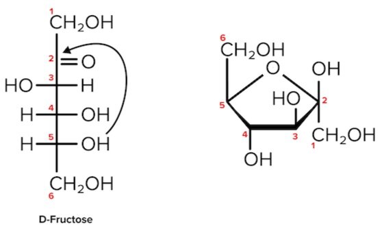
Disaccharides form when two monosaccharides undergo a dehydration reaction. During this process, the hydroxyl group of one monosaccharide combines with the hydrogen of another monosaccharide, releasing a molecule of water and forming a covalent bond. A covalent bond formed between a carbohydrate molecule and another molecule (in this case, between two monosaccharides) is known as a glycosidic bond. In some cases, it’s important to know which carbons on the two sugar rings are connected by a glycosidic bond. Each carbon atom in a monosaccharide is given a number, starting with the terminal carbon closest to the carbonyl group (when the sugar is in its linear form). This numbering is shown for glucose and fructose, above. In a sucrose molecule, the 1 carbon of glucose is connected to the 2 carbon of fructose, so this bond is called a 1-2 glycosidic linkage. Glycosidic bonds are also categorized based on whether the monosaccharide is in the α form or β form .
Common disaccharides include maltose, lactose, and sucrose, as shown below. These disaccharides differ from one another in their monosaccharide constituents and the specific type of glycosidic linkages connecting them. Maltose, or malt sugar, is a reducing sugar joined in an α-1-4-glycosidic linkage, meaning that an α-linkage is made between the first carbon atom of one glucose molecule to the fourth carbon atom of the second glucose molecule. Lactose is a reducing sugar made from β-galactose and α/β-glucose joined by a β-1-4-glycosidic bond. It is found naturally in milk. Sucrose, or table sugar, is a nonreducing sugar made from α-glucose and β-fructose joined at the hydroxyl groups on the anomeric carbons. It is unique among the common disaccharides in having an α-1,β-2-glycosidic linkage.
Key Points
• Disaccharides form when two monosaccharides undergo a dehydration reaction to form a covalent bond known as a glycosidic bond.
• Maltose is composed of two molecules of glucose joined by an α-1,4-glycosidic linkage. It is a reducing sugar that is found in sprouting grain.
• Lactose is composed of a molecule of galactose joined to a molecule of glucose by a β-1,4-glycosidic linkage. It is a reducing sugar that is found in milk.
• Sucrose is composed of a molecule of glucose joined to a molecule of fructose by an α-1,β-2-glycosidic linkage. It is a nonreducing sugar that is found in sugar cane and sugar beets.
Key Terms
Disaccharides: A carbohydrate consisting of two monosaccharides connected to one another through a glycosidic linkage.
Monosaccharides: The basic unit of carbohydrates that cannot be hydrolyzed to simpler chemical compounds with the general formula (CH2O)n.
Glycosidic bonds (linkages): A type of covalent bond that joins two carbohydrate molecules.
Dehydration reaction: A chemical reaction in which two molecules are covalently linked in a reaction that generates a water molecule as a second product.
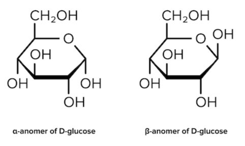
α form : The configuration of a cyclic monosaccharide where the oxygen attached to the anomeric carbon is on the opposite side of the ring from the CH₂ OH group.
β form : The configuration of a cyclic monosaccharide where the oxygen attached to the anomeric carbon is on the same side of the ring as the CH₂ OH group.
Maltose: A disaccharide composed of two glucose molecules connected through an α-1-4-glycosidic linkage.
Lactose: A disaccharide composed of β-galactose and α/β-glucose connected through a β-1-4-glycosidic bond.
Sucrose: A disaccharide composed of α-glucose and β-fructose connected through an α-1,β-2-glycosidic linkage.
α-1-4 glycosidic linkages: Linkages in which carbon atoms 1 and 4 of the two monomers form a glycosidic bond.
β-1-4 glycosidic bonds: Linkages in which carbon atoms 1 and 4 of two glucose monomers in their β form connect through a glycosidic bond.
α-1,β-2-glycosidic linkage: Linkages in which carbon atoms 1 and 2 of an α-glucose and β-fructose connect through a glycosidic bond.
Carbohydrates for the MCAT: Everything You Need to Know
Learn key MCAT concepts about carbohydrates, plus practice questions and answers

(Note: This guide is part of our MCAT Biochemistry series.)
Part 1: Introduction
Part 2: Useful reactions
a) Fischer and Haworth projections
b) Cyclization
Part 3: Sugar structures
a) High-yield monosaccharides
b) High-yield disaccharides
c) Glycogen
Part 4: High-yield terms
Part 5: Passage-based questions and answers
Part 6: Standalone questions and answers
Part 1: Introduction
Although modern humans have been around for at least 100,000 years, one topic that seems to puzzle us to this day is our diet. In grocery stores all across the United States, you’ll find plenty of food products marketed as “low-carb” or “keto-friendly.” You may have even heard of famous athletes, such as Lebron James and Tim Tebow, sharing their results on a low-carb diet.
Despite the bad press, carbohydrates certainly played—and continue to play—a significant role in our evolutionary history and shaped who we are today. Thus, it’s no surprise that you are expected to understand them in detail as you pursue a career in medicine. In this article, we’ll discuss the basic biochemistry behind carbohydrates so that you can have a strong foundation to build on when reviewing their metabolism.
Without further ado, let’s begin!
Part 2: Useful reactions
a) Fischer and Haworth projections
Carbohydrates, also commonly referred to as “sugars,” can be represented in many forms. One such form is a Fischer projection, a two-dimensional representation of a molecule that gives three-dimensional information. The horizontal lines of a Fischer projection are equivalent to wedges: functional groups on horizontal lines are coming out of the page. The vertical lines are equivalent to dashes: functional groups on vertical lines are going into the page.

For sugars, the absolute configuration is designated using D- and L- nomenclature instead of the R and S system used in organic chemistry. If the hydroxyl group of the highest-numbered chiral carbon is on the right, the sugar is in the D-configuration. If the hydroxyl group of the highest-numbered chiral carbon is on the left, it is in the L-configuration.
While Fischer projections represent the straight-chain form of carbohydrates, you may also see sugars represented in their cyclic form as Haworth projections. In a cyclic sugar, the anomeric carbon is the carbon that has two bonds to oxygen.
Be aware that a cyclic sugar can exist in two possible anomers: an ⍺-anomer and a β-anomer. A sugar is in its α-anomer form when the anomeric carbon’s substituent is on the opposite side of the plane as the highest numbered chiral center’s substituent. A sugar is in its β-anomer form when the anomeric carbon’s substituent is on the same side of the plane as the highest numbered chiral center’s substituent.

b) Cyclization
Cyclic sugars are formed via an intramolecular reaction, or a reaction between two functional groups on the same molecule. Aldoses are sugars with an aldehyde group (-CHO). Their cyclization forms a hemiacetal. On the other hand, Ketoses have a ketone group (-CO) and no other further oxidized functional groups. Their cyclization forms a hemiketal.
It’s easiest to visualize this reaction when the carbohydrate is drawn in its linear form (e.g., in a Fischer projection). During cyclization of both aldoses and ketoses, the hydroxyl group (nucleophile) on the highest-numbered chiral center attacks the carbonyl group (electrophile).

Note that as we convert the structure of the sugar from its two-dimensional to three-dimensional structure, the functional group that is pointing to the right of the Fischer projection will end up pointing downward in the Haworth projection.
For most sugars that are dissolved in a solution, this cyclization occurs spontaneously. As a result, many sugars can be found in various combinations of D-, L-, and linear forms. (The final ratios of each of these forms are usually affected by stereochemistry unique to each molecule.) This tendency to undergo spontaneous cyclization is known as mutarotation.
Sugars can also be described as being “non-reducing” or “reducing.” A reducing sugar is one that can act as a reducing agent. Reducing sugars can be identified through the presence of a free anomeric carbon, meaning it is not in a glycosidic bond and has a free hydroxyl group. Conversely, non-reducing sugars lack a free anomeric carbon.
Benedict’s reagent and Tollen’s reagent are two reagents that are commonly used to detect the presence of reducing sugars. The two reagents react with reducing functional groups in unique ways: Benedict’s reagent reacts with aldoses to form a red copper precipitate, while Tollen’s reagent reacts with aldehydes to form a silver, mirrorlike precipitate.


